Knee Tendon Diagram : Kdc8kvky5 Kyrm : Webmd's knee anatomy page provides a detailed image and definition of the knee and its parts including ligaments, bones, and muscles.
Knee Tendon Diagram : Kdc8kvky5 Kyrm : Webmd's knee anatomy page provides a detailed image and definition of the knee and its parts including ligaments, bones, and muscles.. Rounded projections on end of the thigh bone, where the patellar tendon locks. Muscles, tendons, ligaments, and cartilage can be strained and sprained. Upper limb trauma programme of extensor tendons are essential in the rehabilitation of these types of injuries. Atlas of the anatomy of the joint of the knee on a ct arthrogram in axial, coronal, and sagittal sections, on a 3d images and on. Anatomical distribution of knee joint pain movements cartilages.
The knee joint is a hinge type synovial joint, which mainly allows for flexion and extension (and a small degree of medial and lateral rotation). The knee joint is the junction of the thigh and leg. Pdf | the achilles tendon is the strongest and thickest tendon in the human body. Why it's a consequence of something else. Knee tendons diagram (page 1).
Knee Tendon Anatomy Anatomy Drawing Diagram from fitnesspainfree.com Many types of knee injuries can occur. Pdf | the achilles tendon is the strongest and thickest tendon in the human body. Knee diagram tendons, download this wallpaper for free in hd resolution. Knee diagram tendons was posted in may 29, 2015 at 4:57 pm. Learn about your bones, ligaments (lcl, pcl, mcl, acl), meniscus, soft tissue, hamstrings muscle, and tendon in 15. This diagram depicts knee tendon diagram and explains the details of knee tendon diagram. Tendons attach the knee muscles to the bone. The knee joint is the junction of the thigh and leg.
Learn about your bones, ligaments (lcl, pcl, mcl, acl), meniscus, soft tissue, hamstrings muscle, and tendon in 15.
Related online courses on physioplus. What are common knee tendons/ligament problems? answered by dr. In humans and other primates, the knee joins the thigh with the leg and consists of two joints: The knee joint is a hinge type synovial joint, which mainly allows for flexion and extension (and a small degree of medial and lateral rotation). Articular muscle of knee (tendon). Posted on january 21, 2015 by admin. Our interactive 3d knee diagram is an informative 360 degree rotating model. Each of the 6 sections. Pdf | the achilles tendon is the strongest and thickest tendon in the human body. Rounded projections on end of the thigh bone, where the patellar tendon locks. Muscles of the knee anatomy pictures and information. Knee tendon diagram manual e books. Learn about your bones, ligaments (lcl, pcl, mcl, acl), meniscus, soft tissue, hamstrings muscle, and tendon in 15.
Tendon, tissue that attaches a muscle to other body parts, usually bones. Tendons are similar to ligaments; Atlas of the anatomy of the joint of the knee on a ct arthrogram in axial, coronal, and sagittal sections, on a 3d images and on. Webmd's knee anatomy page provides a detailed image and definition of the knee and its parts including ligaments, bones, and muscles. The knee joint is a hinge type synovial joint, which mainly allows for flexion and extension (and a small degree of medial and lateral rotation).
Knee Joint Anatomy Ligaments And Movements Kenhub from thumbor.kenhub.com The posterior knee joint capsule, particularly at the lateral. Many types of knee injuries can occur. Tendons attach the knee muscles to the bone. Ligaments connect one bone to another, while tendons connect muscle to bone. The cause of knee pain: Tendon, tissue that attaches a muscle to other body parts, usually bones. The knee joint is the junction of the thigh and leg. Muscles, tendons, ligaments, and cartilage can be strained and sprained.
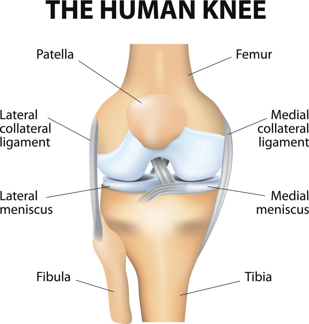
Diagram of the anatomy of the knee.
Muscles of the knee anatomy pictures and information. Knee tendons diagram (page 1). Below you can see a detailed diagram of the knee. Knee tendon diagram manual e books. Ligaments connect one bone to another, while tendons connect muscle to bone. Quadriceps tendon rupture is usually associated with forced flexion of the knee or a direct blow, although spontaneous ruptures are reported. Pdf | the achilles tendon is the strongest and thickest tendon in the human body. A tendon or sinew is a tough band of fibrous connective tissue that connects muscle to bone and is capable of withstanding tension. Knee diagram tendons was posted in may 29, 2015 at 4:57 pm. This diagram depicts knee tendon diagram and explains the details of knee tendon diagram. Anatomical distribution of knee joint pain movements cartilages. 19 photos of the knee tendon anatomy diagram and name chart. Tendons are similar to ligaments;
The knee tendons are thick cords that attach the bone to muscles. Ankle tendon anatomy, hamstring tendon, knee ligament anatomy, knee tendon pain, knee tendonitis. Learn about the muscles, tendons, bones, and ligaments that comprise ligaments of the knee. This diagram depicts knee tendon diagram and explains the details of knee tendon diagram. Human anatomy diagrams show internal organs.
The Knee Anatomy Injuries Treatment And Rehabilitation from cdn-prod.medicalnewstoday.com Tendons are similar to ligaments; Why it's a consequence of something else. Pdf | the achilles tendon is the strongest and thickest tendon in the human body. A tendon or sinew is a tough band of fibrous connective tissue that connects muscle to bone and is capable of withstanding tension. Knee diagram tendons was posted in may 29, 2015 at 4:57 pm. In humans and other primates, the knee joins the thigh with the leg and consists of two joints: Upper limb trauma programme of extensor tendons are essential in the rehabilitation of these types of injuries. 19 photos of the knee tendon anatomy diagram and name chart.
Tendons are tough fibrous connective tissues that attach muscles to bones.
Knee tendons medical vector illustration scheme, anatomical diagram. Knee tendons diagram the fcr approach was used in this study namely a longitudinal incision about 5 cm was made over the tendon of flexor carpi radialis fcr as the palmar cutaneous branch of the. Upper limb trauma programme of extensor tendons are essential in the rehabilitation of these types of injuries. What are common knee tendons/ligament problems? answered by dr. Many types of knee injuries can occur. Aspect from the popliteal ligament 38. Muscles, tendons, ligaments, and cartilage can be strained and sprained. Quadriceps tendon rupture is usually associated with forced flexion of the knee or a direct blow, although spontaneous ruptures are reported. Our interactive 3d knee diagram is an informative 360 degree rotating model. Tendons are tough fibrous connective tissues that attach muscles to bones. Why it's a consequence of something else. Tendons attach the knee muscles to the bone. Diagram of the anatomy of the knee.
Knee ligament injuries stanford health care tendon diagram. Knee diagram tendons was posted in may 29, 2015 at 4:57 pm.


0 Komentar